
A new class of artificial intelligence now maps protein interactions at single-cell resolution. The approach links molecular structure, expression context, and cellular state, predicting which proteins bind or influence each other inside individual cells. This capability narrows the gap between molecular predictions and physiological relevance, enabling drug discovery teams to gain speed, precision, and confidence.
Mapping interactions within single cells is crucial because context shapes biology. The same proteins can behave differently across cell types and states. Tumor cells, immune cells, and neurons, for example, assemble distinct complexes. Bulk assays average these signals, hiding crucial variation. Single-cell models surface actionable details that were previously obscured.
Why Protein Interactions Drive Therapeutic Progress
Most diseases involve disrupted protein interaction networks. Mutations alter binding surfaces or signaling assemblies, and pathogens hijack host complexes to propagate infection. Drugs often work by stabilizing or disrupting specific protein partnerships. Better interaction maps, therefore, guide target selection and mechanism design.
Traditional methods measure interactions in vitro or in bulk tissues. Co-immunoprecipitation, yeast two-hybrid, and affinity purification reveal candidates. These approaches remain essential but miss context. They can overlook transient, weak, or state-dependent contacts. Single-cell inference complements experiments by prioritizing conditions and cell types for validation.
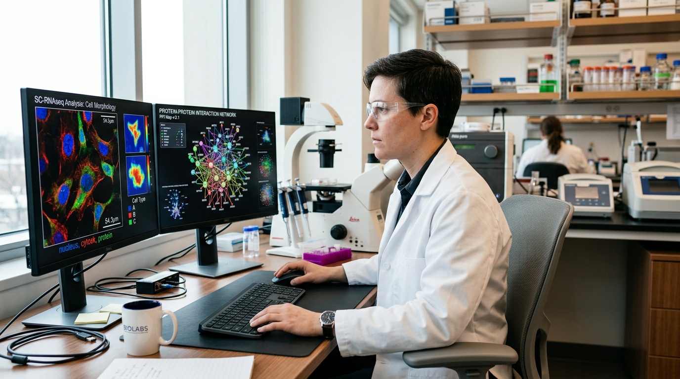
How the AI Model Builds Single-Cell Interaction Maps
The model integrates three information streams. It learns from curated interaction databases containing positive examples. It ingests single-cell omics that describe cellular state. It uses structural and sequence features that affect binding specificity. This multimodal fusion grounds predictions in biology and physics.
Graph neural networks encode molecular networks and propagate neighborhood context. Transformers capture sequence motifs and long-range dependencies. Contrastive objectives align single-cell states with interaction likelihoods. Calibration layers convert raw scores into probabilities. The system outputs interaction probabilities for each protein pair within each cell.
Structural priors strengthen specificity when available. Tools like AlphaFold 3 predict complex structures and interfaces. Docking scores and interface conservation inform compatibility. The model can weigh these features alongside expression. It then downranks pairs that are structurally implausible in a given context.
Training Data and Label Strategy
The model learns from interaction resources such as BioGRID, IntAct, STRING, and HuRI. These databases contain experimentally supported contacts. The team constructs negative pairs using distance and annotation constraints. Hard negatives help the network learn discriminatory patterns. Careful sampling reduces bias and class imbalance.
Single-cell inputs come from public and proprietary datasets. Sources include well-annotated atlases and disease cohorts. The pipeline harmonizes data across sequencing platforms and labs. Normalization, batch correction, and quality control remain essential. Cell-type labels and embeddings anchor predictions to biology.
Representing Cellular Context
The model encodes cellular context using embeddings learned from single-cell RNA-seq. It can optionally include ATAC-seq and proteomic tags. Spatial transcriptomics adds neighborhood and tissue architecture. Cytokine exposure and perturbation metadata refine state definitions. These features allow dynamic, state-specific interaction predictions.
Importantly, the model predicts cell-specific probabilities, not universal truths. A pair may interact strongly in activated T cells. The same pair may remain silent in resting fibroblasts. These differences drive precision pharmacology opportunities. The system highlights where and when biology matters most.
Key Technical Advantages Over Bulk Interaction Maps
Single-cell resolution preserves heterogeneity across tissues and patients. The approach distinguishes rare subpopulations that drive disease. It also captures transitional states during differentiation or treatment. Temporal sampling further reveals dynamic rewiring. These layers expand the interpretability of interaction networks.
Multimodal fusion increases predictive fidelity. Sequence and structure features support molecular plausibility. Expression and chromatin features support cellular feasibility. Spatial features support microenvironment compatibility. Together, these signals reduce false positives and false negatives.
Scalability also differentiates the platform. Efficient architectures handle millions of cells and thousands of proteins. Distributed training addresses memory constraints and data volume. Caching and indexing accelerate inference on new datasets. Engineers design the system for routine screening workflows. This practicality speeds adoption in discovery pipelines.
Accelerating Drug Discovery With Context-Aware Interaction Maps
Target discovery benefits first. The model identifies interactions unique to disease-driving cells. Researchers can prioritize nodes essential for pathological states. These nodes often make better therapeutic targets. They also reduce risk of widespread side effects.
Mechanism of action studies gain clarity. Predicted interaction rewiring explains observed transcriptional or phenotypic changes. Teams can link a compound to disrupted complexes. They can also propose compensatory pathways that restore signaling. This insight refines medicinal chemistry objectives.
Faster Hit Triage and Lead Optimization
Screening yields many hits with uncertain relevance. The model ranks hits by their impact on disease-specific interactions. Chemists focus on compounds that modulate validated complexes. Early triage saves time and resources. It also reduces attrition during later experiments.
Structure-informed predictions aid binding specificity improvements. The system suggests interface residues important for selectivity. These suggestions guide rational modifications. Teams can evaluate predicted off-target interactions efficiently. Safer leads emerge earlier in the pipeline.
Safety, Off-Target, and Polypharmacology Assessment
Safety assessment requires context-aware predictions. The model flags interactions likely in sensitive tissues. It also highlights off-target complexes in cardiomyocytes or neurons. This information complements safety pharmacology and toxicology testing. Early warnings help redesign molecules before costly studies.
Polypharmacology can be beneficial when controlled. The model proposes multi-target profiles aligned with network fragility. Combinations can selectively collapse disease networks. Meanwhile, healthy networks remain buffered. These strategies support rational combination therapy design.
Precision Medicine and Biomarker Development
Patient stratification enhances the success rate of clinical trials. The model facilitates the identification of patient subgroups characterized by targetable interaction signatures. Biomarker panels can monitor network modulation during treatment, enabling clinicians to adapt therapy based on early responses. Adaptive strategies minimize patient exposure to ineffective treatment regimens.
The approach also informs drug repurposing. It scans for compounds that normalize disease-specific interactions. Approved drugs receive priority due to known safety. Rapid translation becomes viable for urgent indications. This acceleration matters during outbreaks and rare disease crises.
Experimental Validation and Benchmarking Practices
Predictions require careful validation. Proximity labeling methods, including BioID and APEX, test interactions in living cells. Proximity ligation assays confirm endogenous interactions. Crosslinking mass spectrometry maps interfaces at scale. Co-immunoprecipitation provides orthogonal evidence under defined conditions.
Functional assays assess phenotypic consequences. CRISPR perturbations test network robustness in targeted cells. Chemical probes evaluate complex stabilization or disruption. Single-cell readouts measure downstream signaling shifts. These assays link predictions to therapeutic relevance.
Benchmarking employs held-out proteins, cell types, and laboratories. Metrics encompass AUROC, AUPRC, and precision at top ranks. Calibration curves ensure probabilities accurately reflect reality. External challenges assess generalization to novel tissues. Transparency and reproducibility are core performance objectives.
Limitations, Risks, and Open Questions
Data quality and coverage still limit performance. Many interactions remain unobserved or context dependent. Negative labels are often uncertain. Class imbalance challenges training and evaluation. Ongoing curation and active learning help address these gaps.
Correlation does not guarantee causation. Co-expression can mimic functional contact without binding. The model may overfit cell-type labels. Experimental design and perturbation tests remain essential. Teams must separate association from mechanism.
Temporal dynamics pose another challenge. Interactions can be transient and stimulus dependent. Snapshot data may miss rapid switches. Time-course experiments and live-cell imaging help. Integrating real-time modalities will strengthen future models.
Finally, computational costs can be substantial. Training across tissues and species demands resources. Efficient architectures and sparse evaluation reduce load. Cloud platforms and accelerators enable scaling. Cost-awareness keeps research accessible and sustainable.
Responsible Deployment and Data Governance
Responsible deployment requires rigorous documentation. Model cards should describe training data, biases, and intended uses. Versioning ensures traceability across studies. Access controls protect sensitive patient information. FAIR principles support reuse and transparency.
Clinical translation adds regulatory obligations. Validation must reflect diverse populations and settings. Auditable pipelines strengthen confidence among regulators. Shared benchmarks reduce duplicated effort. Collaboration accelerates safe adoption across the field.
What Comes Next for AI-Guided Interactomics
Future models will unify structure prediction, dynamics, and single-cell context. Foundation models can learn universal biochemical representations. Active learning will target experiments that reduce uncertainty quickly. Closed-loop automation will tighten discovery cycles, multiplying practical impact.
Integration with spatial proteomics will deepen tissue relevance. Multiplexed imaging and mass spectrometry provide complementary constraints, allowing the model to refine predictions using physical colocalization and prioritize microenvironments for targeted delivery. Precision rises as context becomes richer.
Partnerships between computational and experimental teams will be decisive, fostering shared design, make, test, and analyze loops. Teams will rapidly iterate from interaction maps to chemical series, with feedback sharpening models and hypotheses. The result is faster, smarter therapeutic development.
This new model class reframes protein interaction studies, honoring biological context while exploiting computational power. It transforms complex single-cell data into actionable maps, enabling drug discovery to gain speed, selectivity, and confidence, ultimately benefiting patients with targeted, safer therapies.